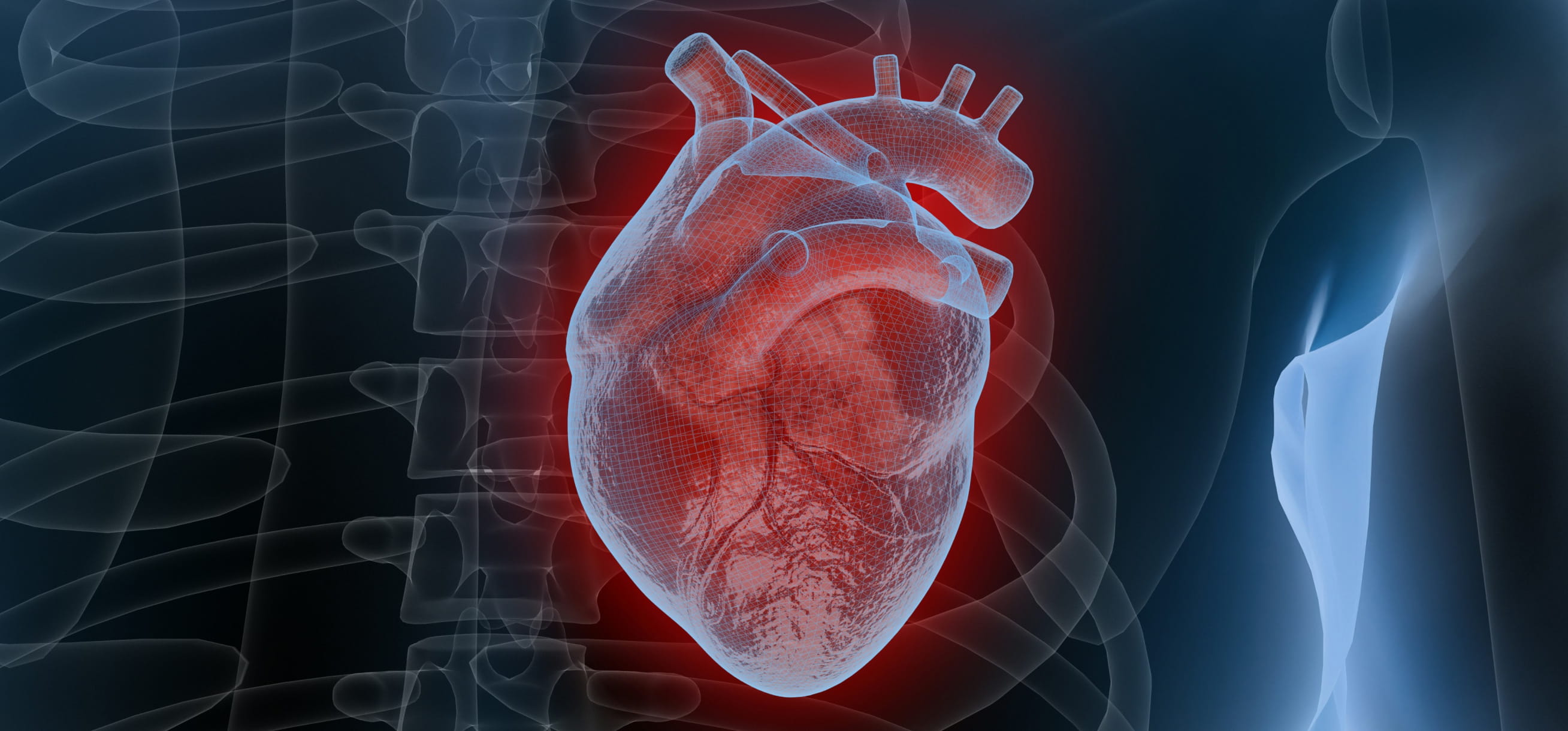
A healthy heart does not beat consistently like a metronome that measures musical beats per minute consistently. The intervals between successive heartbeats vary subtly from one beat to the next, expanding and contracting in response to breathing, movement, stress, digestion, and dozens of other physiological signals. This variation is called heart rate variability, or HRV.
It reflects the dynamic balance between the sympathetic nervous system (which accelerates the heart in response to challenge) and the parasympathetic nervous system (which slows it during recovery and rest).
A heart that can shift fluidly between these two states is a heart that is functioning well. A heart whose beat-to-beat timing is rigid and metronome-like is a heart whose autonomic regulation has become impaired, and that impairment, the science now shows clearly, predicts a wide range of adverse health outcomes including cardiovascular mortality, diabetes, depression, and accelerated biological ageing. For people in Northern Europe HRV is one of the most information-dense biomarkers that can be tracked through a standard consumer wearable.
That is because people in countries like Germany and Finland are burdened by Europe's highest metabolic disease rates, long winter seasons that chronically stress the autonomic nervous system, and a growing epidemic of overwork and sleep insufficiency.
Heart rate variability is the biological variation in the time interval between consecutive heartbeats, quantified in milliseconds. It is regulated by the autonomic nervous system (ANS) and reflects the moment-to-moment competition between sympathetic (arousal) and parasympathetic (recovery) input to the sinoatrial node, the heart's natural pacemaker.
A higher resting HRV generally indicates greater autonomic flexibility and resilience; a lower HRV indicates reduced adaptive capacity and elevated allostatic load.
The evidence base is extensive. A comprehensive meta-analysis of 32 studies and 38,008 participants found that lower HRV was a significant predictor of all-cause and cardiac mortality across all ages, sexes, continents, and recording lengths. [1]
In populations with established cardiovascular disease, the lowest HRV quartile carries a 56% higher hazard ratio for all-cause mortality compared to the top three quartiles. [1] In healthy populations without known CVD, low HRV is associated with a 32–45% increased risk of a first cardiovascular event.[2]
Beyond cardiovascular outcomes, reduced HRV is independently associated with type 2 diabetes and cardiac autonomic neuropathy, [3] depression, anxiety, and PTSD, [4] metabolic syndrome and insulin resistance, [5] and accelerated biological aging.
The metric is also modifiable: aerobic exercise, controlled slow breathing, sleep optimization, and cold water immersion each have documented, measurable effects on resting HRV. [6]
HRV is a real-time window into the health trajectory of your cardiovascular and nervous systems.
The Biology of HRV: What the Gaps Between Beats Measure
The sinoatrial (SA) node generates the electrical impulse that initiates each heartbeat. In a purely mechanical system, each impulse would arrive at precisely regular intervals, a fixed number of beats per minute with no variation.
But the heart is not a closed mechanical system. It receives continuous, competing input from both branches of the autonomic nervous system via direct neural innervation and circulating hormones, and the beat-to-beat timing reflects the outcome of this continuous negotiation.
The Autonomic Nervous System and Cardiac Control
The sympathetic nervous system (SNS) releases noradrenaline onto adrenergic receptors at the SA node, increasing firing rate and reducing inter-beat intervals, the 'fight-or-flight' acceleration. Its effects operate over seconds to minutes. The parasympathetic nervous system (PNS), acting primarily through the vagus nerve, releases acetylcholine, which hyperpolarises the SA node and increases inter-beat intervals: the 'rest-and-digest' slowing.
Parasympathetic effects operate in milliseconds, far faster than sympathetic effects. This means that the rapid, high-frequency variation in inter-beat intervals observed during normal breathing is almost entirely driven by vagal activity. [7]
This is why most HRV metrics particularly RMSSD (root mean square of successive differences), the standard output on consumer wearables, are primarily interpreted as indices of cardiac vagal tone: the strength and responsiveness of parasympathetic, vagus-mediated cardiac control.
The Polyvagal Framework and HRV as Systemic Readiness
The vagus nerve is not merely a cardiac accelerator in reverse. It is the body's primary 'safety' signaling pathway, connecting the brain to the heart, lungs, gut, and immune system. From the perspective of polyvagal theory, and more broadly from the neurovisceral integration model developed by Thayer and colleagues.
A high vagal tone (indexed by high HRV) represents a state of integrated physiological readiness: the organism is not in emergency mode, inflammatory processes are suppressed, the prefrontal cortex can regulate emotional responses, and resources can be directed toward repair, digestion, and immune surveillance. [8]
The prefrontal cortex (PFC) has direct descending projections to the central autonomic network that modulate vagal output. Meaning that cognitive and emotional regulation literally shapes cardiovascular function.
Conversely, reduced HRV reflects a state where the amygdala's threat-detection circuits are dominating, the PFC's regulatory influence is diminished, and the body is in a prolonged state of mobilisation, even when no acute threat is present.
This is the autonomic signature of chronic stress, and it explains why HRV is predictive across such a wide range of diseases: cardiovascular, metabolic, psychiatric, and immunological.
HRV is not a single measurement. It is a family of related metrics, each derived from the sequence of inter-beat intervals (R-R intervals) in a different way. Here is how to understand which metric your device reports, and what it actually captures:
Root mean square of successive differences. Primarily reflects short-term parasympathetic (vagal) activity. The standard output of most consumer wearables (Garmin, Polar, Whoop, Oura, Apple Watch). Most relevant for daily recovery monitoring and stress assessment.
Standard deviation of all normal R-R intervals. Reflects total HRV including both sympathetic and parasympathetic components. Used primarily in clinical 24-hour Holter recordings. SDNN < 50 ms is a strong predictor of cardiovascular mortality; > 100 ms is associated with 5.3× lower mortality risk post-MI.
High-frequency spectral power (0.15–0.4 Hz). Almost entirely vagally mediated. Directly reflects respiratory sinus arrhythmia (RSA) — the increase in heart rate with inhalation and decrease with exhalation. Elevated by slow breathing; reduced by anxiety and chronic stress.
Low-frequency spectral power (0.04–0.15 Hz). Reflects both sympathetic and parasympathetic activity, including baroreflex function. Often interpreted as 'sympathovagal balance' though this remains scientifically contested.
Sympathovagal balance index. Higher values suggest sympathetic dominance. Useful contextually but notoriously variable and not recommended as a standalone daily metric.
Percentage of successive R-R intervals differing by > 50 ms. A simple measure of short-term parasympathetic activity. Correlates strongly with RMSSD and HF power.
HRV declines with age, this is one of the most robust findings in the field.
The Lifelines Cohort Study (n = 84,772 participants) provides the most comprehensive population-based RMSSD reference values to date. [9] Peak RMSSD occurs in adolescence, declines steeply through the second and third decades, and then plateaus after age 60. Women aged 20–45 have meaningfully higher median RMSSD than men (by approximately 5 ms), with the sex difference narrowing after menopause.
A 45-year-old endurance athlete may have an RMSSD above 80 ms; a sedentary 45-year-old with metabolic syndrome may be below 25 ms. Absolute values matter less than trends relative to your own 30-day rolling average.
A sustained drop of ≥ 10% below your rolling baseline is a more actionable signal than any single reading. Lifelong endurance athletes maintain RMSSD values 20–30% higher than age-matched sedentary peers, demonstrating that the age-related decline is substantially modifiable. [10]
The relationship between low HRV and cardiovascular outcomes is the oldest and most extensively validated application of the metric, dating to landmark post-myocardial infarction studies in the 1970s.
The mechanistic logic is clear: impaired autonomic regulation at the cardiac level reduces the heart's ability to respond appropriately to physiological challenge, increasing vulnerability to arrhythmia, ischaemia, and sudden cardiac death.
A meta-analysis of eight studies in 21,988 participants without known CVD found that individuals with the lowest SDNN quintile had a 35% higher relative risk of a first cardiovascular event compared to those with the highest SDNN.
Critically, a 1% increase in SDNN corresponded to approximately a 1% reduction in CVD risk. Suggesting a graded, continuous dose-response relationship. [2] This positions HRV as a potentially useful pre-clinical screening tool, capable of identifying elevated cardiovascular risk years before traditional biomarkers such as elevated LDL or hypertension become abnormal.
In the Multi-Ethnic Study of Atherosclerosis (MESA), borderline abnormal RMSSD (< 5th percentile for age and sex) was associated with significantly increased risk of CVD events and all-cause mortality over an 11-year follow-up, independent of traditional cardiovascular risk factors including age, cholesterol, blood pressure, and smoking status. [12]
In patients with established cardiovascular disease, the prognostic power of HRV increases substantially. A meta-analysis of 28 cohort studies (n = 3,094 CVD patients) found that lower HRV was associated with a 2.27-fold higher risk of all-cause death (HR 2.27, 95% CI: 1.72–3.00) and a 1.41-fold higher risk of cardiovascular events (HR 1.41, 95% CI: 1.16–1.72). [13]
The association was strongest in patients post-acute myocardial infarction, the clinical scenario where HRV measurement is most firmly embedded in guidelines.
In heart failure specifically, a 2024 meta-analysis of 10 studies (n = 10,544 patients) found a pooled effect size of 1.99 for the HRV-mortality association, with SDNN showing the strongest individual prediction. [14] HRV improved risk stratification beyond ejection fraction and NYHA class, the standard clinical metrics, particularly for sudden cardiac death risk. A 2025 synthesis of 67 studies (n = 38,008) reported that SDNN < 70 ms is associated with a 1.5- to 2.3-fold higher risk of major adverse cardiovascular events (MACE). [15]
Cardiovascular disease remains the leading cause of death in both Germany and Finland, accounting for approximately 37% of German deaths and 38% of Finnish deaths annually.
Germany carries one of Europe's highest burdens of metabolic comorbidities, with 11.4% T2D prevalence and a documented north-south gradient of cardiovascular risk across federal states.
Finland's historically elevated cardiovascular mortality (the North Karelia Project was launched there specifically because Finnish CVD rates were among the world's highest in the 1960s) has improved dramatically, but the underlying autonomic and metabolic risk factors persist in a population with high sedentary work rates, winter physical inactivity, and stress-driven sleep disruption.
HRV monitoring in preventive primary care settings in both countries represents an underutilized tool for identifying individuals at elevated autonomic-cardiovascular risk before clinical events occur.
The relationship between HRV and metabolic disease is bidirectional and self-reinforcing.
Chronic sympathetic dominance (low HRV) promotes insulin resistance through multiple pathways:
A systematic review and meta-analysis confirmed strong evidence for overall HRV decrease in type 2 diabetes, with impairment affecting both sympathetic and parasympathetic domains. [3]
Crucially, the ARIC (Atherosclerosis Risk in Communities) study found that autonomic impairment is detectable at early stages of metabolic dysregulation, before clinical diabetes diagnosis.
Diabetic subjects showed a significantly more rapid temporal decline in HRV over a 9-year follow-up compared to normoglycaemic controls (SDNN decline: −0.95 ms/year vs. −0.65 ms/year in non-diabetics). [16] This suggests HRV decline may precede, and potentially predict, progression to frank diabetes, not merely reflect it.
In a Finnish-led study examining HRV and metabolic syndrome components (n = 220 adults, aged 23–70 years, conducted in part by Verve Research, Oulu, Finland), women with metabolic syndrome had significantly lower SDNN and low-frequency HRV power than metabolic-syndrome-free women. [5]
Waist circumference, the abdominal obesity marker, was the metabolic syndrome component most consistently associated with lower HRV. This is clinically relevant for both German and Finnish populations, where abdominal obesity prevalence has increased substantially over the past two decades.
Reduced HRV is associated with insulin resistance through parasympathetic dysfunction, even in non-obese individuals. [17]
The proposed mechanism: sympathetic overactivation suppresses insulin-stimulated glucose uptake in skeletal muscle, while simultaneously reducing insulin sensitivity through cortisol and catecholamine-mediated pathways.
This positions HRV monitoring as a potentially valuable tool for early identification of insulin resistance before standard fasting glucose and HbA1c testing crosses diagnostic thresholds, particularly relevant given that insulin resistance precedes clinical T2D by 5–15 years in most individuals.
Perhaps the most underappreciated dimension of HRV research is its relationship to psychiatric and psychological health. The neurovisceral integration model proposes that the prefrontal cortex's capacity for emotion regulation is directly indexed by vagal tone.
Meaning HRV is not merely a cardiac metric but a window into the brain's ability to regulate its own responses to stress, threat, and social challenge.
An umbrella review of meta-analyses examining HRV across mental disorders found consistent evidence for decreased HRV in major depressive disorder (MDD), with reductions in RMSSD and high-frequency power reflecting diminished parasympathetic activity. [18]
Interestingly, evidence from HRV biofeedback intervention studies suggests the relationship may be causal in both directions: depression suppresses HRV, but interventions that artificially raise HRV (through resonance-frequency breathing) produce measurable improvements in depressive symptoms. [19]
A UK Biobank analysis of 15,768 participants identified distinct autonomic clusters, with one low-HRV pattern specifically associated with greater depression prevalence and suicide attempt rates.
A meta-analysis of 36 studies (2,086 anxiety patients, 2,294 controls) found that anxiety disorders as a class are characterised by significantly reduced resting HRV, with a small-to-moderate effect size (Hedges' g = −0.29 for HF power). [20] The mechanism: chronic threat-biased attention activates corticotropin-releasing factor pathways, chronically withdrawing parasympathetic activity.
The impairment is state-independent, reduced vagal HRV is found even in individuals who score high in trait anxiety without a formal diagnosis, and even during periods of low acute anxiety. This makes resting HRV a potential endophenotypic marker for anxiety vulnerability.
PTSD is characterised by autonomic dysregulation: the threat-response system becomes chronically hyperactivated, producing persistently reduced HRV. A 2024 meta-analysis of HRV biofeedback (HRVB) interventions for PTSD in military veterans found a moderate-to-large effect size (Hedges' g = −0.557, p < 0.001) for symptom reduction, with the additional advantage of markedly lower attrition (5.8%) compared to standard psychological PTSD treatments. [21]
This places HRVB as a promising complementary intervention for trauma, particularly for populations resistant to standard talk therapies.
Finland has clinically significant rates of both depression and PTSD, compounded by seasonal affective disorder (SAD) affecting an estimated 9.5–21% of the population depending on measurement criteria.
The autonomic suppression associated with winter darkness, reduced morning light cortisol response, circadian phase delay, reduced physical activity, maps directly onto the HRV reductions seen in depressive disorders.
This means that for Finnish adults monitoring HRV through the winter, a sustained seasonal decline in RMSSD is not merely a fitness metric: it may represent an early signal of SAD onset, providing an objective anchor for deciding when to intensify morning light therapy, increase exercise volume, or seek clinical support.
HRV declines with chronological age, but the rate of decline is not fixed.
Biological aging, as distinct from calendar aging, proceeds faster in individuals with chronic autonomic dysregulation, chronic inflammation, poor sleep, and sedentary behaviour. In this sense, resting HRV can be understood as a proxy for biological age: a 55-year-old with the HRV of a 35-year-old is, in a functional physiological sense, biologically younger than their birth year implies.
The cardiovascular health study demonstrated that HRV predicts cardiovascular mortality in older adults independent of traditional risk factors including blood pressure, cholesterol, diabetes, and smoking history. [22]
This independent predictive power suggests HRV captures aspects of ageing physiology: autonomic flexibility, inflammatory regulation, mitochondrial function, that conventional biomarker panels miss.
The capacity of lifelong endurance athletes to maintain HRV values 20–30% above age-matched peers [10] demonstrates that the decline is not inevitable: it is, to a substantial degree, a lifestyle disease of the autonomic nervous system.
You can change your HRV. The evidence across multiple intervention modalities is now sufficiently consistent to rank approaches by evidence strength and practical accessibility. The following summarizes the current state of the science.
Endurance aerobic training is the most robustly documented long-term HRV enhancer. A 2024 narrative review of lifelong endurance athletes found sustained RMSSD values 20–30% higher than age-matched non-athletic peers. [10]
The mechanism operates through structural cardiac remodeling (increased vagal efferent tone), reduced resting sympathetic outflow, and improved baroreflex sensitivity.
Practical protocol: 150 minutes per week of zone-2 cardio (conversational-pace aerobic exercise, roughly 60–70% maximum heart rate) can raise RMSSD by approximately 8 ms over 12 weeks. [10]
Strength training at 70–80% of 1RM, performed 2–3 times per week, also enhances vagal tone in middle-aged adults, particularly when combined with aerobic training. [23]
Voluntary slow breathing at approximately 6 breaths per minute (0.1 Hz), sometimes called resonance-frequency or coherence breathing, is the most potent acute HRV enhancer with documented effects within a single session.
A meta-analysis of 223 studies found significant increases in vagally-mediated HRV during slow breathing, immediately after a session, and after a multi-session intervention programme. [24]
The mechanism: at 6 bpm, the breathing cycle aligns with baroreflex oscillations (~0.1 Hz), creating resonance that maximises the amplitude of respiratory sinus arrhythmia and baroreflex gain.
Even 2-minute sessions of slow breathing produce measurable HRV increases.
Sustained multi-week practice produces lasting improvements in resting HRV. [25] HRV biofeedback, slow breathing with real-time visual feedback of the HRV waveform, adds an additional training signal that accelerates learning and produces benefits for anxiety, depression, hypertension, and PTSD symptomatology. [26]
HRV is highest during deep non-REM (slow-wave) sleep and lowest during REM sleep and the pre-waking period. Each additional hour of quality sleep adds approximately 3 ms to overnight RMSSD. [27]
Sleep deprivation below 7 hours is associated with significantly reduced next-day resting HRV, elevated inflammatory markers, and impaired glucose metabolism. A trifecta that compounds over time into metabolic and cardiovascular risk.
For both German and Finnish populations: where work pressure and artificial lighting chronically compress sleep, sleep duration and quality represent the foundational HRV intervention that no other practice can substitute.
Cold water immersion (CWI) after exercise consistently shows positive acute effects on parasympathetic reactivation, as measured by HRV. A systematic review of 12 RCTs found all studies reported parasympathetic reactivation post-CWI, with six demonstrating statistically significant improvements compared to passive recovery. [28] The initial cold shock response produces a brief sympathetic surge (lasting ~20–90 seconds), followed by a parasympathetic rebound that restores and often exceeds baseline HRV. For Nordic populations — particularly Finnish users culturally familiar with cold immersion through sauna-followed-by-cold-plunge traditions. This represents a well-tolerated, culturally integrated HRV intervention with strong mechanistic support.
RCT evidence supports modest RMSSD increases (~3–4 ms). Anti-inflammatory mechanism reduces sympathetic tone. Particularly relevant in Finland where fatty fish consumption is culturally embedded.
Consistent RMSSD improvements across multiple RCTs. Effect size moderate (Hedges' g ≈ 0.4–0.6). The Wim Hof Method (breathwork + cold) showed superior momentary HRV improvements vs. meditation-only in a 29-day RCT (n=404).
Cofactor for parasympathetic neurotransmission and vagal signalling. Deficiency is common in Germany and Finland (estimated 30–40% of European adults). Supplementation shows modest HRV benefit, particularly in deficient individuals.
Even moderate alcohol (1–2 drinks) measurably suppresses overnight HRV. Reduction is among the most impactful single dietary changes for resting HRV in habitual drinkers.
Visceral fat is the metabolic syndrome component most strongly associated with low HRV. Weight loss of 5–10% body weight in overweight individuals produces significant HRV improvements.
Short-term studies show measurable HRV increases during and after exposure to natural environments vs. urban environments. Mechanism likely involves reduced sympathetic arousal and sensory stress.
The same factors that drive cardiovascular, metabolic, and psychiatric disease also reduce HRV, a convergence that explains why HRV functions as such a broad-spectrum risk biomarker. The following have documented, quantified suppressive effects on resting HRV:
Chronically elevated cortisol and catecholamines suppress parasympathetic activity; one of the strongest and most consistent HRV suppressors
Each hour of lost sleep reduces next-day RMSSD; chronic sleep debt produces structural autonomic changes
One of the primary mechanisms linking sitting time to cardiovascular risk; independent of exercise volume
Abdominal fat is the metabolic syndrome component most closely linked to reduced HRV; associated with chronic low-grade inflammation and sympathetic overactivation
Progressive autonomic neuropathy reduces both sympathetic and parasympathetic HRV components; decline accelerates after diagnosis
Suppresses overnight HRV even at 1–2 drinks; dose-dependent effect; disrupts sleep architecture
Directly damages autonomic nerve fibres and reduces vagal tone; independent of cardiovascular structural damage
Chronic sympathetic dominance and reduced vagal tone are core autonomic features of both conditions
Autonomic hyperarousal chronically suppresses HRV; among the most consistent autonomic signatures in trauma literature
Bidirectional: low HRV permits higher inflammation (reduced vagal anti-inflammatory reflex); high inflammation further suppresses vagal tone
Paradoxically, excessive training volume without adequate recovery suppresses HRV; athlete monitoring uses RMSSD trends to detect non-functional overreaching before injury
Single HRV readings are noisy and largely uninterpretable. What matters is trend relative to your own rolling baseline. A 30-day rolling average RMSSD provides a stable reference point. Actionable signals:
HRV Decision Framework
HRV does not exist in isolation. Its value as a health signal is amplified when interpreted alongside complementary laboratory biomarkers that illuminate the pathways driving autonomic dysregulation.
Through the Aniva Health platform, the following testing framework is recommended for individuals with persistently low or declining HRV:
Chronic low-grade inflammation directly suppresses vagal tone through the inflammatory reflex. Elevated hsCRP (> 3 mg/L) is both a driver and consequence of low HRV.
Identifies pre-diabetic metabolic dysregulation that accelerates autonomic neuropathy and HRV decline. HRV decline often precedes glucose abnormalities, testing confirms whether metabolic damage is occurring.
Blunted or exaggerated morning cortisol rise indicates hypothalamic-pituitary-adrenal axis dysregulation — the hormonal counterpart of low HRV. In SAD, the cortisol awakening response is typically suppressed.
Low vitamin D is associated with autonomic dysregulation and reduced vagal tone, particularly relevant in Finland (70% of children vitamin D deficient in winter). Target > 75 nmol/L year-round.
Detects insulin resistance before glucose becomes abnormal. Elevated HOMA-IR (> 2.5) correlates with reduced HRV and identifies metabolic risk at least 5 years before T2D diagnosis.
Thyroid function modulates cardiac autonomic regulation. Both hypo- and hyperthyroidism alter HRV. Essential to rule out thyroid contributions to HRV abnormalities.
Iron deficiency impairs mitochondrial energy production and reduces exercise tolerance, blunting the HRV-raising effect of aerobic training. Correction of iron deficiency may be necessary before HRV responds to exercise interventions.
RBC magnesium reflects intracellular status more accurately than serum. Deficiency impairs parasympathetic neurotransmission. Estimated 30–40% of European adults are deficient.
Directly reflects dietary omega-3 status. Index > 8% is associated with significantly higher HRV and lower cardiovascular risk. Most Europeans are below 6%.
Conclusion
Heart rate variability is unusual among health biomarkers in that it is simultaneously a window into the past (reflecting accumulated autonomic damage from chronic stress, poor sleep, metabolic disease, and sedentary behaviour), a snapshot of the present (reflecting today's recovery status and physiological readiness), and a predictor of the future (forecasting cardiovascular events, metabolic deterioration, and psychiatric risk years before clinical thresholds are crossed).
The evidence base is now sufficiently mature to support three clear conclusions.
References
All references are peer-reviewed publications, systematic reviews, or meta-analyses.
[1] Jarczok MN, et al. (2022). Heart rate variability in the prediction of mortality: A systematic review and meta-analysis of healthy and patient populations. Neuroscience & Biobehavioral Reviews, 143:104907. n = 38,008. PMID: 36243195
[2] Hillebrand S, et al. (2013). Heart rate variability and first cardiovascular event in populations without known cardiovascular disease: meta-analysis and dose-response meta-regression. EP Europace, 15(5):742–749.
[3] Vanderlei LCM, et al. (2018). Heart rate variability in type 2 diabetes mellitus: a systematic review and meta-analysis. PLOS ONE. doi:10.1371/journal.pone.0195166
[4] Wang Z, et al. (2025). Heart rate variability in mental disorders: an umbrella review of meta-analyses. Translational Psychiatry. doi:10.1038/s41398-025-03339-x
[5] Stuckey MI, et al. (2015). Associations between heart rate variability, metabolic syndrome risk factors, and insulin resistance. Applied Physiology, Nutrition, and Metabolism, 40(7):734–740. Oulu, Finland cohort.
[6] Laborde S, et al. (2022). Effects of voluntary slow breathing on heart rate and heart rate variability: a systematic review and meta-analysis. Neuroscience & Biobehavioral Reviews, 138:104710.
[7] Task Force of the European Society of Cardiology and the North American Society of Pacing and Electrophysiology. (1996). Heart rate variability: standards of measurement, physiological interpretation and clinical use. Circulation, 93(5):1043–1065.
[8] Thayer JF, Lane RD. (2007). The role of vagal function in the risk for cardiovascular disease and mortality. Biological Psychology, 74(2):224–242.
[9] Tegegne BS, et al. (2020). Reference values of heart rate variability from 10-second resting electrocardiograms: the Lifelines Cohort Study. n = 84,772 participants. PMC7734556.
[10] Damoun N, et al. (2024). Heart rate variability and endurance athletes: HRV in lifelong athletes 20–30% above age-matched peers. Narrative review. Applied Physiology, Nutrition, and Metabolism.
[11] Stone JD, et al. (2021). Wearable technology real-world validation of photoplethysmography-derived RMSSD. International Journal of Sports Physiology and Performance.
[12] Shah AJ, et al. (2016). Reference ranges for short-term heart rate variability in individuals free of cardiovascular disease: MESA. n = 6,332 validation cohort. PMC5010946.
[13] Fang SC, et al. (2020). Heart rate variability and risk of all-cause death and cardiovascular events in patients with cardiovascular disease: a meta-analysis of cohort studies. n = 3,094. PMID: 31558032
[14] Yadav R, et al. (2025). Heart rate variability as a predictor of mortality in heart failure: a systematic review and meta-analysis. Cureus, 17(12):e99120. n = 10,544. PMC12794729.
[15] Addleman S, et al. (2025). Reduced resting HRV and risk of major adverse cardiovascular events: synthesis of 67 studies (n = 38,008). Applied Psychophysiology and Biofeedback.
[16] Carnethon MR, et al. (2003). Diabetes, glucose, insulin, and heart rate variability: the Atherosclerosis Risk in Communities (ARIC) study. Diabetes Care, 28(3):668–674.
[17] Toon Health Study (2015). Heart rate variability, insulin resistance, and insulin sensitivity in Japanese adults. PMC4549610.
[18] Bassett D, et al. (2024). The predictive potential of heart rate variability for depression. Psychiatry Research. doi:10.1016/j.psychres.2024.00126X
[19] Chalmers JA, et al. (2014). Anxiety disorders are associated with reduced heart rate variability: a meta-analysis. Frontiers in Psychiatry. PMC4092363.
[20] Tomasi J, et al. (2024). Heart rate variability: evaluating a potential biomarker of anxiety disorders. Psychophysiology. doi:10.1111/psyp.14481
[21] Steele AL, et al. (2024). Heart rate variability biofeedback for PTSD in military veterans: meta-analysis. Military Medicine, 189(9-10):e1903.
[22] Stein PK, et al. Novel measures of heart rate variability predict cardiovascular mortality in older adults independent of traditional cardiovascular risk factors: The Cardiovascular Health Study.
[23] Lin LLC, et al. (2022). High-intensity resistance training and HRV in middle-aged adults. International Journal of Environmental Research and Public Health.
[24] Laborde S, et al. (2022). ibid. Systematic review and meta-analysis of slow breathing on HRV. n = 223 studies. Neuroscience & Biobehavioral Reviews.
[25] Pozzato I, et al. (2025). Single 2-minute slow breathing sessions and HRV increases. Respiratory Physiology & Neurobiology.
[26] Lehrer P, et al. (2020). Heart rate variability biofeedback improves emotional and physical health and performance: a systematic review and meta-analysis. Applied Psychophysiology and Biofeedback.
[27] Nicolini P, et al. (2024). Sleep duration and HRV: each additional hour of quality sleep adds approximately 3 ms to overnight RMSSD. Sleep Medicine.
[28] Galvez-Rodriguez T, et al. (2025). Cold water immersion and parasympathetic HRV reactivation post-exercise: systematic review of 12 RCTs. European Journal of Applied Physiology.